Menu
Menu

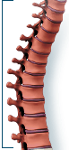

Spinal Fusion Surgery
Spinal fusion is a surgical procedure that involves placing bone graft material between adjacent vertebrae to promote bone growth that joins together, or "fuses," the two structures. In patients for whom it’s appropriate, the procedure may be performed using minimally invasive surgical techniques.
What is Spinal Fusion Surgery?
Spinal fusion is a surgical technique in which one or more of the vertebrae of the spine are joined together (fused) to stop them from moving against each other. This is done by placing bone grafts or bone graft substitutes between the affected vertebral bone. The graft material acts as a binding medium and also helps to maintain normal disc height – as the body heals, the vertebral bone and bone graft eventually grow together to join the vertebrae and stabilize the spine.
Why Do I Need This Procedure?
There are a number of reasons your surgeon may recommend spinal fusion. This procedure is frequently used to treat
- One or more fractured (broken) vertebrae
- Spondylolisthesis (slippage of one vertebral bone over another)
- Abnormal curvatures of the spine, such as scoliosis or kyphosis
- Protruding or degenerated discs (the cartilaginous “cushions” between vertebrae)
- Instability of the spine (abnormal or excessive motion between two or more vertebrae)
Spinal fusion is typically recommended only after conservative treatment methods fail. Your surgeon will take a number of factors into consideration before making this recommendation, including the condition to be treated, your age, health and lifestyle and your anticipated level of activity following surgery. If you are considering spinal fusion, please discuss this treatment option thoroughly with your spinal care provider.
How Is Spinal Fusion Performed?
There are a variety of surgical approaches and procedures, but all involve the placement of bone graft material between vertebrae. The graft material may be bone – either taken from the patient (autograft) or from a bone bank (allograft) – or a synthetic bone substitute called bone morphogenetic protein (BMP).
The spine may be approached and the graft positioned either from the back (Posterior Lumbar Interbody Fusion [PLIF]), the front (Anterior Lumbar Interbody Fusion [ALIF]) or the side (Tranforaminal Lumbar Interbody Fusion [TLIF]). Your surgeon also may decide that more than one approach is necessary. Please discuss your fusion approach options thoroughly with your surgeon,and rely on his or her judgment about which is most appropriate for your particular condition.
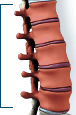
Instrumentation, such as screws, plates and cages, may be used to create an “internal cast” to support the vertebral structure during the healing process.
Spinal Fusion: Traditional vs. Modern Approach
Traditionally, surgeons have performed spinal fusion as an open procedure, which involves making an incision, stripping bands of muscle and retracting muscle and tissue for a clear view of the spine and easy access to the vertebrae for implantation.
Traditionally, autograft has been “the gold standard” in graft material. However, removal of the bone – usually from the patient’s pelvis or iliac crest – can be very painful. Allograft does not require this extra procedure, but healing often is not as predictable as with the patient’s own bone. BMP, a genetically produced protein, prompts the patient’s own bone cells to make more bone.
Modern spinal fusion can employ less invasive surgical techniques, such as muscle dilation, making the highly invasive posterior fusion approach unnecessary in many cases.
Muscle dilation is achieved by using a series of sequential dilators, or tubes to separate the fibers of the back muscles and create a small tunnel, enabling the surgeon to view the spine through an incision less than an inch long and leaving the muscle virtually intact. Advances in instrumentation allow rods and screws to be inserted via tiny incisions in the skin.
How Long Will It Take Me To Recover?
The recovery period for spinal fusion will vary depending on the procedure and your body’s ability to heal and firmly fuse the vertebrae together.
Patients typically stay in the hospital for several days, longer if necessary for more extensive surgery. This may also include time in a rehabilitation unit. Your surgeon will prescribe pain medication as needed, and may recommend a brace and follow-up physical therapy.
The length of time you will be off work will depend on a number of factors: your particular fusion procedure and the physician’s approach to your spine, the size of your incision, and whether or not you experienced any significant tissue damage or complications. Another consideration is the type of work you plan to return to. Typically, you can expect to be on medical leave for 3 to 6 weeks.
Work closely with your spinal surgeon to determine the appropriate recovery protocol for you, and follow his or her instructions to optimize the healing process.
Are There Any Potential Risks Or Complications?
All treatment and outcome results are specific to the individual patient. Results may vary. Complications such as infection, nerve damage, blood clots, blood loss and bowel and bladder problems, along with complications associated with anesthesia, are some of the potential risks of spinal surgery. A potential risk inherent to spinal fusion is failure of the vertebral bone and graft to properly fuse, a condition that may require additional surgery.
Please consult your physician for a complete list of indications, warnings, precautions, adverse effects, clinical results and other important medical information that pertains to a spinal fusion procedure.
The materials on this Web site are for your general educational information only. Information you read on this Web site cannot replace the relationship that you have with your health care professional. We do not practice medicine or provide medical services or advice as a part of this Web site. You should always talk to your health care professional for diagnosis and treatment.
Lumbar fusions
Spinal fusion, also called arthrodesis, is a surgical technique used to join two or more vertebrae (bones) within the spine. Lumbar fusion technique is the procedure of fusing the vertebrae in lumbar portion of the spine (lower back).
Lumbar fusion surgery may be used to treat, spondylolisthesis (slipping of the spine bones), degenerated discs, scoliosis or kyphosis (abnormal curvature of the spine), spinal infections or tumors, traumatic injury of the spine, recurrent disc herniation, and unstable spine.
The surgery can be done as an open or laparoscopic (keyhole) surgery.
Your surgeon may approach your spine from the back, abdomen, or neck, depending on the area to be treated.
In spinal fusion, a piece of bone, taken from other parts of the body or donated from a bone bank is transplanted between the adjacent vertebrae. Screws, plates, or cages may be used with the bone graft to help hold the spine.
During the surgery, your surgeon performs a discectomy where a portion of the diseased or damaged disc material is removed. After the removal the roof of the vertebra will be trimmed or removed to relieve the pressure on the nerve and this procedure is called as laminectomy. Following laminectomy, the bone graft (small chips of bone) will be placed alongside of the vertebrae between the vertebrae to be fused. Screws are placed into the vertebrae to be fused. Rods are attached to connect the screws, to stabilize and hold the bones together while the fusion heals.
As with every surgery, lumbar fusion surgery is associated with certain complications and they include:
- Spine infection
- Damage to the spinal nerves
- Loss of sensation
- Problems with bowel or bladder control
- Dislocation of the implant
- Pseudarthrosis, a painful condition occurring as a result of non healing of the bone effusion, and a false joint grows at the site
- Blood clot formation in the legs
- Pain at the bone graft site
Other Lumbar (Low Back) List
- ALIF – Anterior Lumbar Interbody Fusion
- Lumbar Laminectomy
- Lumbar Microdiscectomy
- TLIF – Transforaminal Lumbar Interbody Fusion
- Balloon Kyphoplasty
- Minimally Invasive Lumbar Fusion
- Minimally Invasive Microdiscectomy (METRx Discectomy)
- Deformity correction- Adult
- X-Stop
- Lumbar Total Disc Replacement
- Recovery